
Supplement Protocol
Pre WO
Intra WO
Post WO
Let’s talk about eccentric training and nutrition. Too often we bifurcate nutrition and training as it is a good thing. The detachment of what we are doing peri-workout nutrition or overall nutrition along with peri-workout supplementation and overall supplementation is easier to manage but could be a huge missed opportunity.
One area I am focussed on is increasing my Deficit in Strength Deficit. I was 1.05.
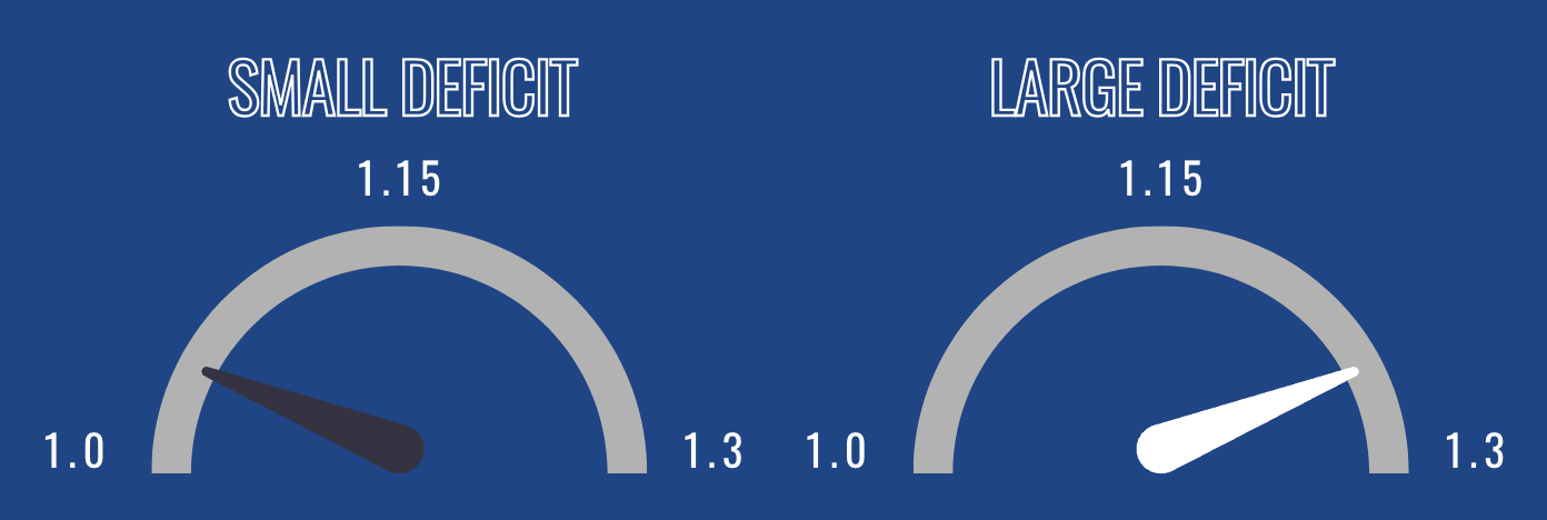
Learn more about Strength Deficit Here: Strength Deficit: Leveraging Ratios Between Eccentric Versus Concentric
So this means I will do a ton of eccentric focus. We have a lot of ways we can with this. One is yielding isometrics, slow eccentrics, overloaded eccentrics, and last but not least fast eccentrics. Each method has a slightly different impact, but the majority is centered around two key concepts.
- Increasing fascicle length and longitudinal hypertrophy
- Increase connective tissue function to support the stretch-shortening cycle
From an architectural perspective, we know that eccentric training changes both muscular tissue and connective to react better to stretch. That is the foundation of increasing the deficit – have a shorter amortization from an eccentric load. If there is no difference between a counter-movement jump and a non-counter-movement jump, there is a limitation coming from the eccentric load of the tissues. The simplest way to break down this is we are incurring acute injury to our connective tissue and muscular tissue every time we train. When adhering to an eccentric focus training block, we should approach supplementation as if we were healing from a grade 1 strain. The faster we can regenerate tissue, the better we can perform the following day.
Sidebar about training. I am an advocate of block orientation and saturating a quality. This does a couple of things, it isolates the stress and allows for more transparency in regards to adaptive potential. For the block I was going through I did a breakdown on the pH Membership site You can sign up here: pH Membership
Lets break down some research on why I focussed on Phosphatidic Acid, Collagen, and BPC 157.
Collagen
- Effects of Collagen Peptides on Recovery Following Eccentric Exercise in Resistance-Trained Males-A Pilot Study
- Resistance-trained males consumed 15 g/day of Collagen (n = 7) or placebo (n = 8), and after 7 days, maximal voluntary isometric contraction (MVIC), countermovement jump height, soreness, and collagen turnover were examined. Five sets of 20 drop jumps were performed and outcome measures were collected 24, 48, and 120 hr postexercise.
- Countermovement jump height was maintained in the Collagen group at 24 hr (PRE = 39.9 ± 8.8 cm vs. 24 hr = 37.9 ± 8.9 cm, p = .102), whereas the Collagen group experienced a significant decline at 24 hr (PRE = 40.4 ± 7.9 cm vs. 24 hr = 35.5 ± 6.4 cm, p = .001; d = 0.32).
- In both groups, muscle soreness was significantly higher than PRE at 24 hr (p = .001) and 48 hr (p = .018) but not at 120 hr (p > .05). MVIC in both legs showed a significant time effect (left: p = .007; right: p = .010) over the 5-day postexercise period.
- CP supplementation attenuated performance decline 24 hr following muscle damage. Acute consumption of CP may provide a performance benefit the day following a bout of damaging exercise in resistance-trained males.
- Influence of specific collagen peptides and 12-week concurrent training on recovery-related biomechanical characteristics following exercise-induced muscle damage—A randomized controlled trial
- 75 subjects were enrolled Part of the pre-test and post-test were 150 drop jumps (6 sets of 25 repetitions, 2 min rest between sets) from a 60 cm (23.5 “) high box. Participants were required to land slowly and in a controlled manner at a knee angle of at least 90° to force an adequate eccentric movement and subsequently induce muscle damage, as described elsewhere
- Training Intervention (12 Weeks) subjects were instructed to perform bodyweight-based lower-body exercises (squats, lunges and calf raises) three times a week, with at least 1 day off between two training days. Weeks 1–4 required 3 sets of 20, weeks 5–8 required 3 sets of 25, and weeks 9–12 required 3 sets of 30 repetitions for each exercise 3x per week.
- Once a day for 12 weeks, they were required to take 15 g of either a placebo (Silicea, metabolically inert) or the test product [mixture of specific collagen peptides (10 g PeptENDURE® + 5 g TENDOFORTE®
- the main findings of the study indicate a higher muscular regenerative capacity in subjects supplementing specific collagen peptides (SCP) compared to PLA. This is reflected in significant improvements in the biomechanical parameters maximum voluntary contraction (MVC) and rate of force development (RFD), which recovered significantly faster at each measurement time point (post, 24 and 48 h), and countermovement jump height (CMJ) at 48 h following 150 drop jumps in the SCP group. To the authors’ knowledge, this is the first study to examine the effects of daily SCP supplementation combined with a 12-week training intervention on parameters associated with muscular recovery.
- They concluded that ECM remodeling occurred more rapidly in the passive/elastic tissues in the CP group, resulting in improved force output and power production.
- resulted in a similar CMJ height after 24 h and a more pronounced but non-significant recovery after 48 h in the CP group compared to whey protein supplementation
- improvement in reactive strength (CMJ height) following SCP administration one and 2 days after muscle-damaging exercise. Higher stiffness results in more efficient use of elastic energy but is reduced during fatigue states (
- Maximal strength has been shown to be moderately related to reactive strength
- The shorter the interval, the smaller the deviation from MVC and thus from muscle size (48) which is why a window of 0–50 ms and 0–100 ms (for peak RFD) was chosen to determine whether SCP affects neural-dependent mechanisms (49). SCP supplementation had a pronounced positive effect on RFD, corroborated by a significant 3-way interaction effect.
- They found a significant correlation between stiffness and RFD100 ms, which describes 30% of the variance in RFD in a sample size of 15 subjects. However, caution should be exercised when attributing stiffness to RFD because inter-individual variability is higher than that of trainability
- CP has been used successfully to significantly reduce pain during activity after 12 weeks and 5 g of CP daily (53, 54).
-
- In the present study, participants reported their perceived muscle soreness after all tests were performed at each time point. Activity-related pain would have impacted the outcome of CMJ, a high range-of-motion exercise.
- To date, satellite cells, inflammatory cells, fibro-adipogenic-precursors, vascular tissue, nerve-muscle interaction and the ECM appear to play a central role, not only in the recovery of functionality but also in tissue adaptation to new mechanical stimuli
- Neural and musculotendinous adaptations, remodeling of the ECM, and inflammation likely regulate RBE (63). Some factors have already been influenced by CP supplementation and therefore represent a promising approach to improving RBE
-
Phosphatidic Acid
- Efficacy of phosphatidic acid ingestion on lean body mass, muscle thickness and strength gains in resistance-trained men
- Sixteen resistance-trained men were randomly assigned to a group that either consumed 750 mg of PA (n = 7, 23.1 ± 4.4 y; 176.7 ± 6.7 cm; 86.5 ± 21.2 kg) or a placebo (PL, n = 9, 22.5 ± 2.0 y; 179.8 ± 5.4 cm; 89.4 ± 13.6 kg) group. During each testing session subjects were assessed for strength (one repetition maximum [1-RM] bench press and squat) and body composition. Muscle thickness and pennation angle were also measured in the vastus lateralis of the subject’s dominant leg.
- All groups performed the same 4-day per week, split routine resistance training program for 8-weeks The load for the assistance exercises was self-determined by the subject, but they were required to use a load that allowed them to perform a 10–12 RM. A 90-s rest period was required between each set, for all exercises.
- One of the signaling proteins that PA has been suggested to stimulate is mammalian target of rapamycin (mTOR) [8,9], a serine threonine kinase that integrates metabolic signals from various factors including protein metabolism and cytoskeleton organization that controls cell growth
- mechanical activation from external loads (as one may see from a resistance exercise session) may be enhanced with the presence of PA
- It has been shown that exogenous supplied PA can stimulate the mTOR pathway via its activation of the substrate S6 kinase
- Subjects ingesting PA demonstrated a 12.7% increase in squat strength and a 2.6% increase in LBM, while subjects consuming PL showed a 9.3% improvement in squat strength and a 0.1% change in LBM.
- Since PA is thought to act on a parallel pathway to a protein stimulus (specifically leucine) on activating the mTOR pathway [8,27], the greater benefit towards increases in lower body strength and lean body mass in PA suggests that the ingestion of this supplement may enhance lean tissue accruement and lower body strength to a greater extent than protein supplementation or resistance exercise only.
- The role of phospholipase D and phosphatidic acid in the mechanical activation of mTOR signaling in skeletal muscle
- elevation in PA concentration was sufficient for the activation of mTOR signaling. Second, the isozymes of PLD (PLD1 and PLD2) are localized to the z-band in skeletal muscle (a critical site of mechanical force transmission). Third, mechanical stimulation of skeletal muscle with intermittent passive stretch ex vivo induced PLD activation, PA accumulation, and mTOR signaling. Finally, pharmacological inhibition of PLD blocked the mechanically induced increase in PA and the activation of mTOR signaling.
- Furthermore, we showed that mTOR signaling was partially resistant to rapamycin in muscles subjected to mechanical stimulation. Because rapamycin and PA compete for binding to the FRB domain on mTOR, these results suggest that mechanical stimuli activate mTOR signaling through an enhanced binding of PA to the FRB domain on mTOR.
- These results indicate that an elevation in [PA] leads to an activation of mTOR signaling in skeletal muscle.
- In summary, the results of this study indicate that mechanical stimuli activate mTOR signaling through a PLD-dependent increase in [PA]. Because the activation of mTOR signaling is required for mechanically induced growth of skeletal muscle, these findings contribute significantly to our understanding of how mechanical signals are transduced into the molecular events that regulate skeletal muscle growth and remodeling.
- Phosphatidic acid mediates activation of mTORC1 through the ERK signaling pathway
- The mammalian target of rapamycin (mTOR) assembles into two distinct multiprotein complexes known as mTORC1 and mTORC2. Of the two complexes, mTORC1 acts to integrate a variety of positive and negative signals to downstream targets that regulate cell growth. The lipid second messenger, phosphatidic acid (PA), represents one positive input to mTORC1, and it is thought to act by binding directly to mTOR, thereby enhancing the protein kinase activity of mTORC1. Support for this model includes findings that PA binds directly to mTOR and addition of PA to the medium of cells in culture results in activation of mTORC1.
- rather than acting directly on mTORC1, the results suggest that exogenous PA must be metabolized to lysophosphatidic acid (LPA), which subsequently activates the LPA receptor endothelial differentiation gene (EDG-2).
- leucine does not act through phospholipase D and PA to activate mTORC1 and, instead, show that the two mediators act through parallel upstream signaling pathways to activate mTORC1.
- results demonstrate that leucine and PA signal through parallel pathways to activate mTORC1 and that PA mediates its effect through the ERK pathway, rather than through direct binding to mTOR.
- Phosphatidic acid: a novel mechanical mechanism for how resistance exercise activates mTORC1 signalling
- In any event, the finding by O’Neil and colleagues (O’Neil et al. 2009) that mechanical stimulation of mTORC1 is dependent on the synthesis of phosphatidic acid is exciting and an excellent addition in our effort to elucidate the complex pathways through which resistance exercise regulates mTORC1 signalling and promotes muscle growth
- The role of phosphoinositide 3-kinase and phosphatidic acid in the regulation of mammalian target of rapamycin following eccentric contractions
- On the contrary, ECs induced a prolonged activation of mTOR (>12 h) and the synthesis of PA by PLD was determined to be necessary for mTOR activation
- he results from this study also demonstrate that PA can activate mTOR through a PI3K–PKB-independent mechanism
- Finally, it was determined that an increase in [PA] and activation of mTOR does not occur in response to all types of contractions, but instead appears to be specific to growth promoting contractions such as ECs
BPC 157
- BPC 157’s effect on healing
- Elements thought to be of greatest importance in the process of healing are formation of granulation tissue, angiogenesis and production of collagen.
- In our work we tested the influence of BPC 157 on: granulation tissue and collagen formation, on angiogenesis as well as on tensile strength development, using three experimental rat models: 1) skin incisional wounds; 2) colon-colon anastomoses; and 3) angiogenesis model with synthetic sponge implantation.
- It is worth noting that these effects were achieved by different routes of application, including intragastric and local, making BPC 157 a potentially useful therapeutic agent.
- Pentadecapeptide BPC 157 (PL 14736) improves ligament healing in the rat
- stable gastric pentadecapeptide BPC 157 (GEPPPGKPADDAGLV, an anti-ulcer peptide effective in inflammatory bowel disease therapy (PL 14736)) particularly improved healing of transected tendon and muscle and wound healing effect including the expression of the early growth response 1 (egr-1) gene.
- BPC 157 was effective given per-orally (0.16 µg/ml in the drinking water (12 ml/day/rat)) until sacrifice. Commonly, BPC 157 µg-ng-rats exhibited consistent functional, biomechanical, macroscopic and histological healing improvements. Thus, we suggest BPC 157 improved healing of acute ligament injuries in further ligament therapy.
- Novel Therapeutic Effects in Rat Spinal Cord Injuries: Recovery of the Definitive and Early Spinal Cord Injury by the Administration of Pentadecapeptide BPC 157 Therapy
- we showed the particular points of the immediate effect of the BPC 157 therapy that began rapidly after its administration, (i) soon after injury (10 min), or (ii) later (4 days), in the rats with a definitive spinal cord injury.
- BPC 157 rats presented only discrete edema and minimal hemorrhage and increased Nos1, Nos2, and Nos3 values (30 min post-injury, (i)) or only mild hemorrhage, and only discrete vacuolation of tissue (day 4, (ii)). In the day 4–30 post-injury study (iii), BPC 157 rats rapidly presented tail function recovery, and no demyelination process (Luxol fast blue staining).